Categories
The rectum and anus form the final part of the digestive tract – the colon. In this relatively small part of the intestine a number of different diseases occur. The diseases in anus and rectum are:
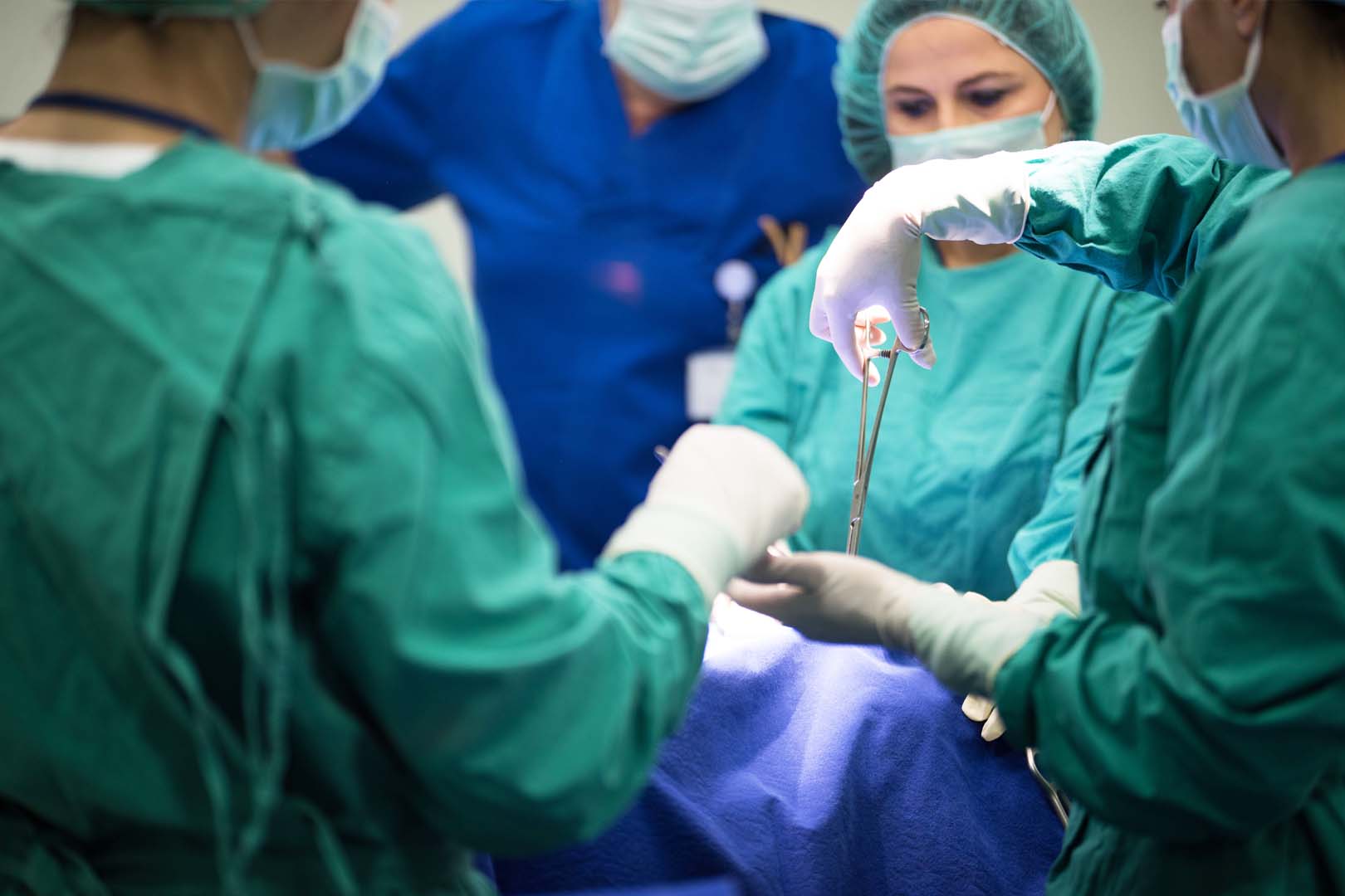
Injuries in anus and rectum and the presence of a foreign body – most of these injuries occur accidentally after a sharp object falls as a result of therapeutic manipulation, insertion of foreign bodies by sexual arousal, etc. The clinical picture varies, depending on the size of the foreign body, how long it has been retained in the rectum, possible inflammation or perforation of the intestine. The diagnosis is made with a rectal douching and a rectal examination. The treatment is surgical depending on the severity of the injury. The type of anesthesia is usually spinal.
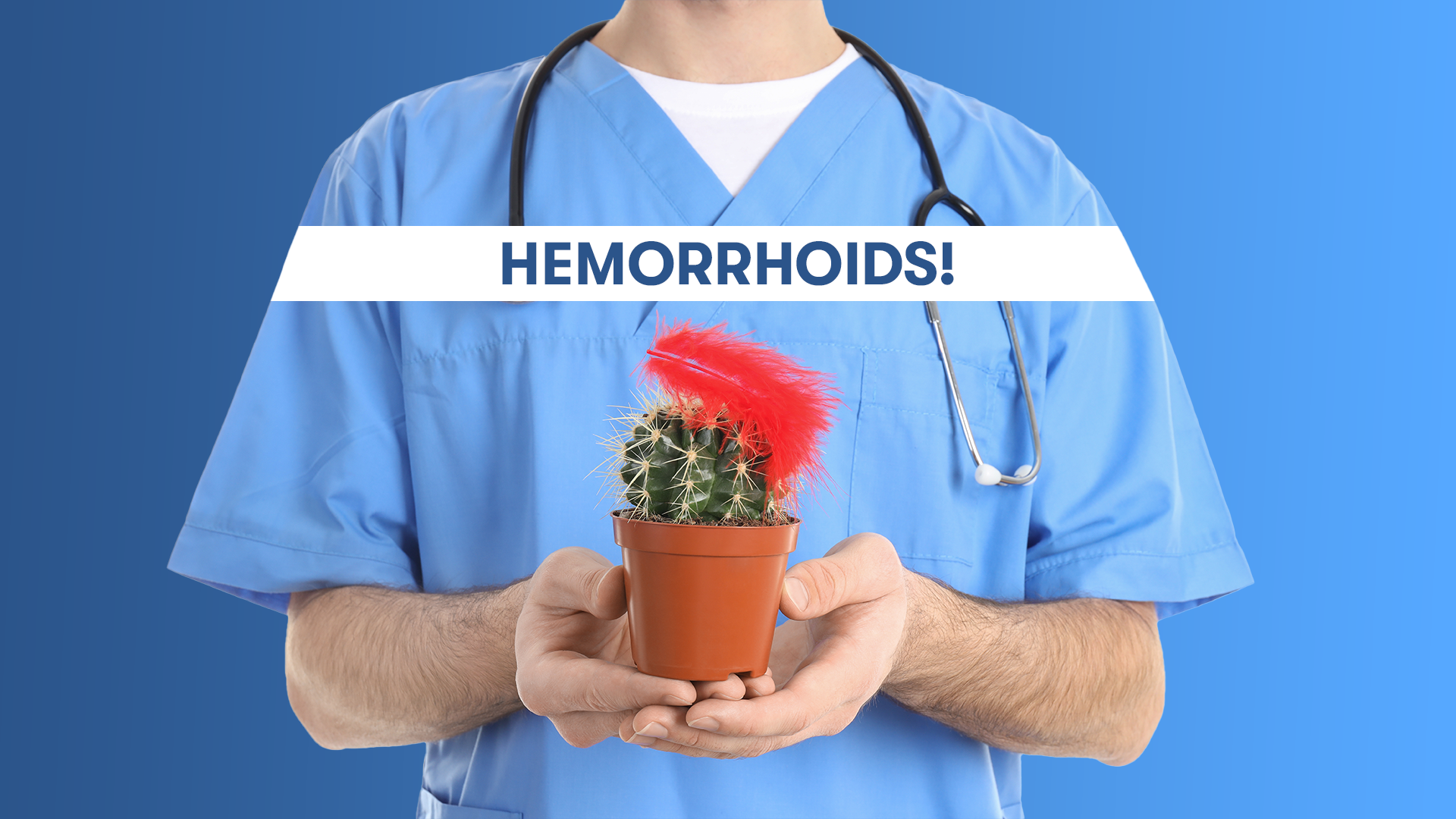
Hemorrhoidal disease – is a pathological enlargement of the hemorrhoidal venous ganglion, upper and lower, or popularly known as internal and external hemorrhoids. Symptoms of this disease are: pain in the defecation, swelling in and around the gum, presence of pure red blood at the end of the defecation, palpable knots around the gum. The causes of this disease are multifactorial – hereditary component, weakness of the vein wall muscle, inflammatory processes in that region, pregnancy, occupations associated with prolonged sitting, internal organs disease, etc. The diagnosis of the disease is based on an external examination of the anus or endoscopically. Depending on how advanced the disease is we distinguish 4 stages. The treatment may be conservative or surgical, depending on the stage of the disease.
Absorption of the anorectal region – occurs as a result of invasion of the pararectal spaces by pathogenic microorganisms and occurs more frequently in men. Most often the cause of the infection begins in anal crypts. The patient complains of pain that occurs when sitting or moving, which becomes more severe over time, followed by elevated body temperature. A diagnosis by a surgeon is sufficient to make a diagnosis. If spontaneous emptying of the pus does not occur, surgery should be performed, with incision and drainage. The intervention is done in a septic room and in spinal anesthesia. A common late complication of this disease is the development of ano-rectal fistula.
Anal fissure – This disease is an ulcer of the anal mucosa that occurs most often in patients who have difficulty emptying and straining during defecation. The clinical picture is manifested by extremely severe pain that is described as scarring, burning. The characteristic of anal fissures is their chronicity with periods of deterioration and improvement. A careful rectal examination is sufficient to diagnose this disease. The early treatment is conservative and, in cases of chronicity – surgical and performed in spinal anesthesia.
Anorexic fissure – occur as a consequence of inflammatory processes in that area. They can be simple or complicated. The disease is usually chronic in nature. The presence of chronic purulent secretion through the paranasal opening is important for the diagnosis of this disease. Sometimes it is necessary to perform an X-ray examination with contrast or fistulography. The treatment is exclusively surgical and in spinal anesthesia. Worldwide, the recurrence or recurrence of the disease is high.
A mastectomy is a surgical removal of the breast. Which type of mastectomy will be used is a decision of the surgeon who performs the operation in agreement with the oncologist and the plastic surgeon if in one operation the breast is reconstructed. The most common cause is breast cancer in women and less often but not excluded in men. It is performed under general anesthesia and in some cases, in addition to removing the breast, it is necessary to remove the lymph nodes. Mastectomy can be simple mastectomy, modified radical, radical, mastectomy with preservation of the skin, mastectomy with preservation of the nipple, prolonged radical mastectomy.
This procedure involves placing a catheter through the urethra to the bladder. In certain medical conditions and in certain procedures it is necessary to place a catheter to measure the amount of urine, to pay attention to the appearance and concentration, but sometimes to remove the accumulated urine in the bladder for some reason that caused a stop. The procedure is performed by a doctor and it is painless. If catheterization is done and should be continued at home, then training for proper manipulation is done. Catheter removal is also a procedure performed by a doctor in a hospital setting.
Gallstones in the gallbladder occur by accumulating its contents, cholesterol and bilirubin. The range in size is from very small (1-2mm) like sand, to very large that fill the entire gallbladder and have its shape. Often the patient has a mixture of all sizes.
The gallstones in the gallbladder are present in 20-30% of the population, are more common in females, the ratio is 4:1. In 80% of cases they are made of cholesterol and the rest of bilirubin. In some cases, small gallstones give a picture of bile sludge and never form a stone. Many patients have no symptoms, in which case it is “silent” or asymptomatic calculosis.
In other “unfortunate” patients the symptoms are: pain under the right costal arch and scoop and may spread to the right shoulder, nausea, vomiting. In some cases, with mild calculus, a pebble can penetrate the main gallbladder, obstructing bile leakage, and in this case the first symptom in the patient is yellowing of the skin and visible mucous membranes, followed by elevated body temperature and white and oily manure. In this case it is a complication of the disease.
The standard method for proving biliary calculus is abdominal ultrasound. In some cases, when the surgeon assesses, more complex diagnostic tests – ERCP and MRCP – may be necessary.
Cholelithiasis is unfortunately only a surgical disease (treated only by surgery). The standard method of operative treatment worldwide is the Laparoscopic method, or popularly known as ‘four-hole surgery’. The type of anesthesia is always with general anesthesia. With this operation, the patient has no incision as before, excluding the possibility of ventral hernias at the site of surgery, the duration of surgery is shortened, the need for antibiotics and pain medication is reduced, the amount of anesthesia is reduced, postoperative pain is drastically reduced, the patient rapidly mobilizes and rapidly switches to fluid and food intake by mouth. The rehabilitation of the patient is much faster, and the working ability is possible after one week. At the same time, the laparoscopic method allows examination of other organs in the stomach and possibly their operative treatment, i.e. “two operations in one”’.
Choledocholithiasis is the presence of pebbles in the main bile duct that carries the bile from the gall bladder to the duodenum. This condition causes the bile to leak into the duodenum and return to the liver causing inflammation and the possibility of liver damage with serious complications.
The symptoms of this disease are:
– Severe pain under the costal arch
– Nausea and vomiting, especially after a fatty meal
– Yellow staining on skin and mucous membrane (icterus)
– The stools become white and greasy and resembles clay
– Increased body temperature with fever, fatigue, in case of inflammatory process of bile ducts (cholangitis)
The diagnosis of this disease is made on the basis of anamnesis and objective examination, laboratory studies, abdominal ultrasound, ERCP and MRCP. Treating this disease can be conservative and surgical.
If within the ERCP the papillotomy is done and pebble removed, it would be the best. It belongs to the field of interventional internal medicine.
The surgical treatment is done under general anesthesia, can be laparoscopic (less often) or classic. This basically removes the gallbladder, opens the main gallbladder, removes the pebble or pebbles from it, thoroughly checks for any residual pebbles present in it. The surgery eventually ends with a special tube being inserted into the canal which is removed after two weeks of surgery. Earlier bile duct contrast imaging checks the bile tract.
Hernia, or popularly known as “kila “or “bruh”, is a combination of increased intra-abdominal pressure (when lifting heavy objects, constipation, prolonged coughing and sneezing, tumors, heart failure, etc.) and muscle or facial openness or weakness. The increased pressure pushes the organs and tissues through the weak spot on the wall.
Muscle weakness can be congenital, but it usually occurs at a later time in life. Depending on the place of occurrence we distinguish: Ingenual, Hemoral, Umbilical, Epigastric and extremely rare Spiegel hernia. The special group includes the ventral or incisal sutures, which occur at the site of a previous abdominal surgery.
Hernia knows no age limit, it affects both men and women. It manifests itself as a painless or slightly sensitive swelling of the stomach skin, which usually occurs after physical exertion and disappears when the patient is lying down.
Evolutionarily, as time goes by, the hernia increases in size. Exceptionally at an early age it can happen that the muscle wall develops that the weak spots naturally strengthen. That is why at an early age you can be treated with operative treatment.
Hernia is a disease that is treated exclusively by surgery. Unless there are concomitant diseases in the patient and surgical treatment is at increased risk, then it is advisable to wear seat belts. Usually, surgical interventions on uncomplicated hernias and in a properly prepared patient, i.e. “Cold operations” fall into the low-risk intervention group and the possibility of complications is minimized. In that case, the fear of surgery is unjustified. Otherwise, if the patient has not resolved his problem in time and comes with complicated or ‘stuck’ hernia – this is the case when the contents of the lean body cannot be returned to the abdominal cavity i.e. the ‘hot’ operation has to be done the risk and the possibility of complications increase. Therefore the recommendations are for earlier surgical treatment.
Surgical treatment of hernias can be by open, laparoscopic or mixed methods. The type of anesthesia is regional or spinal anesthesia and the hospital treatment lasts 24 hours. The modern recommendations that our hospital follows are that every hernia, with rare exceptions, is made with prosthetic material i.e. network. The recovery period and relative sparing of severe physical exertion lasts 3-4 weeks.
Lipoma is a benign formation of adipose tissue, painless, localized on any part of the body, of different size, one or more in number. A physical examination by a surgeon is needed to make a diagnosis. There are several types and rarely this change can be malignant, so to differentiate them the doctor may need additional tests such as echosonographic examination of soft tissues, computed tomography, magnetic resonance imaging, biopsy. No treatment is usually needed. If it is an aesthetic problem for you or the doctor has recommended removal, then the treatment is surgical removal, with minimal extraction and as small a scar as possible, under local anesthesia.
